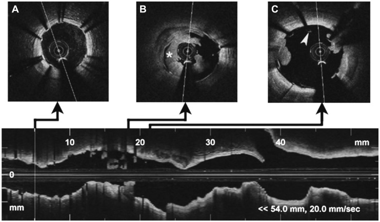
Fig. 3.
Optical coherence tomography imaging of left anterior descending artery. Distal (A): correct apposition of the stent without intraluminal content. Middle (B): a signal rich, low-backscattering protrusion image compatible with white thrombus (mark), which occupies the greater part of the vessel lumen. Proximal (C): stent malapposition (arrow head) in the proximal border, with small images of thrombus.

