
Figure 4.
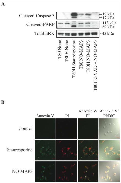
MAP3 NO-donor silica nanoparticles induce apoptosis in ovarian cells. T80 and T80H cells were untreated, treated with 10 μM staurosporine for 1 h, or treated with 400 μg/mL of NO-releasing L-MAP3 nanoparticles alone or in combination with 100 μM z-VAD-FMK for 6 h. Cell lysates were subjected to western blot analysis to probe for the apoptotic markers, cleaved-caspase 3 and cleaved-PARP, or loading control, total ERK1/2 (A). T80H cells were untreated or treated with either 10 μM staurosporine for 1 h or 400 μg/mL of NO-releasing L-MAP3 nanoparticles for 6 h. Cells were then stained with annexin V Alexa Fluor 647 to label apoptotic and PI to label necrotic cells and then imaged under the Zeiss laser scanning confocal microscope (B).