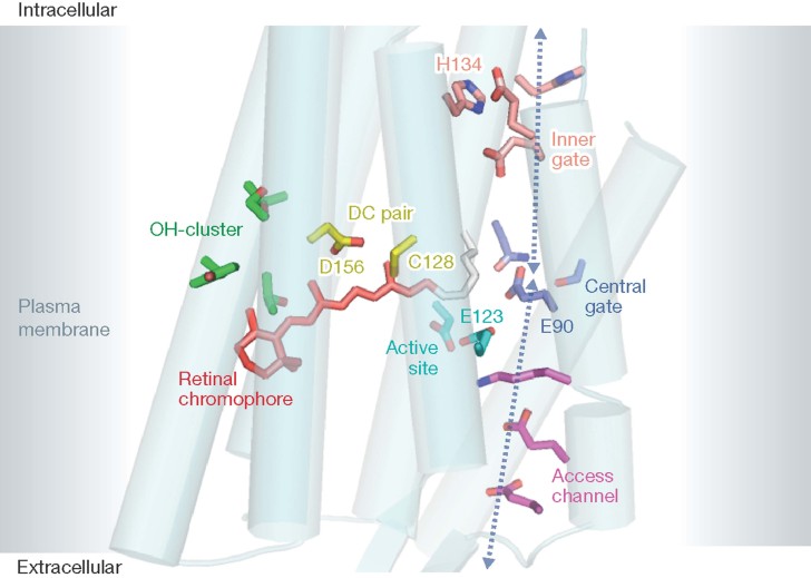
Figure 2. Cartoon of the Channelrhodopsin 7TM-fragment.
The structure is drawn according to the data of Kato et al (2012) with key residues shown in color: voltage sensor E123 (cyan), residues of the access channel (magenta), central gate (blue), and inner gate (orange), OH-cluster green, and the retinal Schiff base is seen in red.

