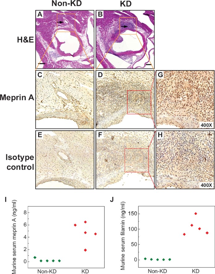
Figure 4. Meprin A is enriched in coronary artery lesions in a mouse model of Kawasaki disease.
- A,B. Micrographs of hematoxylin and eosin-stained sections of the aortic root and coronary arteries of control (A) and LCWE-injected mice (B) demonstrating severe aortitis with intimal proliferation leading to concentric obstruction in the LCWE-injected but not control animals. Arrows point to the normal (A) and diseased (B) coronary arteries.
- C,D. Micrographs of meprin A immunohistochemistry-stained inset areas of the sections of coronary arteries demonstrating enrichment of meprin A in the mononuclear infiltrates of coronary arteries in LCWE-injected (D) but not control (C) animals.
- E,F. Micrographs of isotype control-stained sections of coronary arteries in LCWE-injected (F) and control (E) mice.
- G,H. High-magnification micrographs of the inset areas of meprin A (G) and isotype control-strained (H) sections of the coronary arteries in LCWE-injected mice.
- I,J. Serum levels of meprin A (I) and filamin (J) are elevated in the LCWE-injected (red) as compared to control (green) mice. Scale bar is 250 µM.

