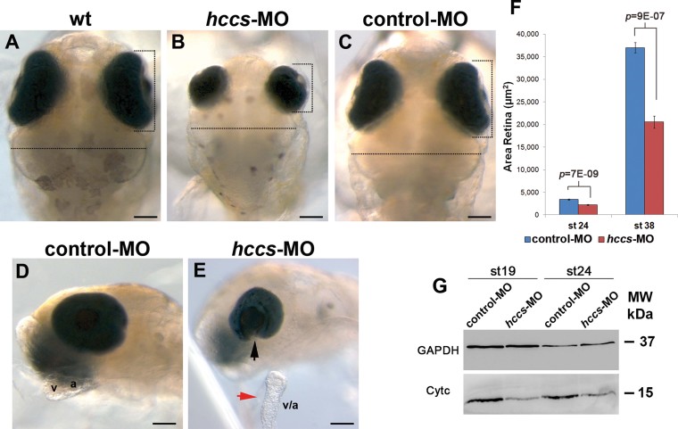
A–E. Bright-field dorsal (
A–C) and lateral (
D,
E) views of wt (
A),
hccs-MO (
B,
E) and control-MO (
C,
D) -injected embryos at st38.
hccs-morphants display microphthalmia (vertical dashed line in
B), microcephaly (horizontal dashed line in
B) and cardiac defects including failure of heart loop formation and pericardial oedema (red arrow in
E). In a significant number of the microphthalmic embryos (50%), RPE layering and closure of ventral optic fissure were also impaired resulting in coloboma formation (black arrow in
E; see also Supporting Information
Table S1). Embryos injected with control-MO did not show any abnormal phenotype (
C,
D). a, atrium; v, ventricle. Scale bars: 100 µm.

