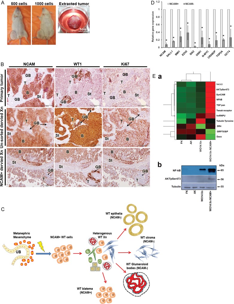
Representative images of NOD-SCID mice injected with 1000 and 500 NCAM+ cells that developed tumours and of a tumour extracted from a mouse injected with NCAM+ cells showing encapsulation of the tumour mass that separate it from host tissues (Scales bars = 100 µm, magnification ×10 and ×20).
Immunohistochemical staining presented in serial sections for NCAM, WT1 and Ki67 in a representative primary tumour and its derived xenografts generated from unsorted cells or from NCAM+ cells showing recapitulation of the original tumour. The NCAM+ cells formed tumours that contain both undifferentiated NCAM+ blastemal structures and well-differentiated NCAM− tubular structures. WT1, the marker used in clinical practice for histopathoplogic diagnosis of WT, is abundantly expressed in both WT xenografts on similar structures as in the primary tumour. Ki67 is more abundant in the WT Xn than in their primary source. T-Tubules; B-Blastema; GB-Glomeruloid bodies; St-Stroma. (Scales bars = 200 µm; Magnification 20×).
A scheme illustrating the putative derivation of NCAM+ WT cells from the transformed MM of the human foetal kidney and their capability to initiate p-WT xenografts that recapitulate the histology of their parental tumour, consisting of NCAM+ blastema cells (possibly formed by self renewal) as well as the differentiated features seen in primary human WTs (immature tubular epithelia, stroma and glomeruloid bodies).
qRT-PCR analysis for the expression of renal progenitor (i.e. NCAM, SALL1, SIX2, OSR1), stemness (i.e. BMI1, EZH2, OCT4) and poor prognostic (i.e. TOP2A, N-MYC, CRAB2P) genes between NCAM+ and NCAM− WT Xn cells (n = 3), demonstrate significantly elevated mRNA levels of these genes in NCAM+ compared to NCAM− cells (p = 0.031). The values for the NCAM+ cells were used to normalize (therefore = 1) and all other values were calculated with respect to them. Results are presented as the mean ± SEM of five separate experiments.
Increased expression of TWEAK-R/TGFb/PKBpSer473/14-3-3/NF-κB specifically in WT xenografts derived from NCAM+ cell. (a) Heat map of Panorama® Antibody Array illustrating expression of differentially expressed proteins in early WT xenograft generated from NCAM+ cells (WO14 Xn NCAM+), WT xenograft generated from cells that were not separated (W014 Xn), human adult kidney (AK) and human foetal kidney (hFK) tissues; (b) Western Blot analysis of NF-kB and AKTpSer473.

