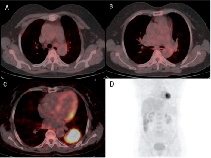
Figure 1.
A 67-year-old woman, with blood CEA of 14.95 ng/ml, was diagnosed with poorly differentiated adenocarcinoma in the inferior lobe of the left lung. Three different slices of integrated FDG-PET/CT images (A–C) and maximum intensity projection showed an SUVmax of the primary tumor of 9.4. Although there was no obvious mediastinal lymph nodal metastasis on imaging, the high SUVmax of the primary tumor and CEA level indicated that the patient might be at risk for lymph nodal metastasis. Pathologic results after surgery showed micro-metastases in subaortic and subcarinal lymph nodes (N2).

