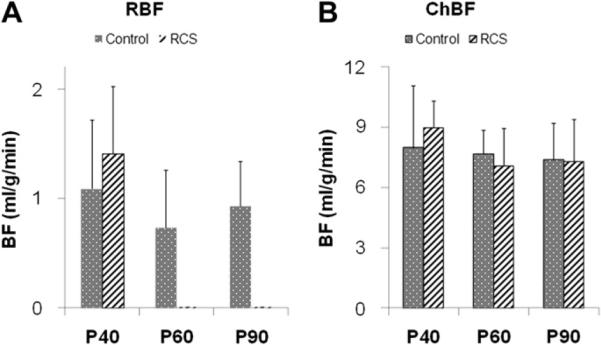
Fig. 5.

Retinal blood flow (RBF) peak values (A) and choroidal blood flow (ChBF) peak values (B) at P40, P60 and P90 of normal and RCS rats (mean ± SD). RBFs became unresolvable at P60 and P90 in the RCS rats. At P40, RBFs were not significantly different between the normal and RCS group (t-test: p = 0.42). RBFs of all three normal groups were not significantly different (ANOVA: p = 0.83). ChBFs were not significantly different at all three time points (t-test: p = 0.53, 0.56 and 0.90, respectively), and ChBFs of all groups were not significantly different (ANOVA: p = 0.63).
