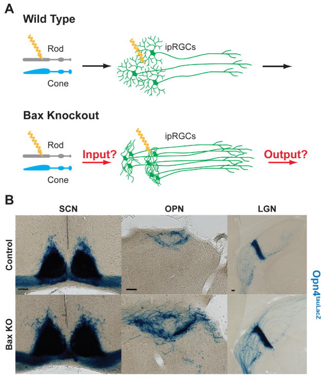
Figure 3. Axonal targeting remains in Bax knockout.
(A) Model describing how lack of apoptosis in ipRGCs might affect reception of light information from rods and cones, and/or transmission of light information to downstream nuclei in the brain. (B) Axons from ipRGCs were labeled by X-gal staining with the Opn4tau-LacZ reporter allele in coronal sections of the SCN (left panel), OPN (middle panel), and IGL (right panel). ipRGC axons still confine to the SCN, OPN, and IGL in the Bax KO similar to the control. (Scale bar 100μm)

