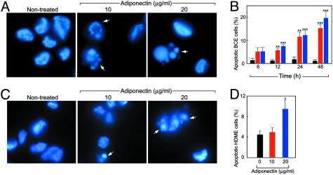
Fig. 3.
Adiponectin induces endothelial apoptosis. (A and B) Apoptotic bodies of adiponectin-treated BCE cells were detected at 24 h by fluorescent microscopy and quantified at different time points. Bars: black, control; red, 10 μg/ml; blue, 20 μg/ml adiponectin. (C and D) Apoptosis of HDME cells after 24-h incubation with adiponectin. Values represent mean percentage of apoptotic cells per total number of cells per field. *, P < 0.05; **, P < 0.01; ***, P < 0.001.

