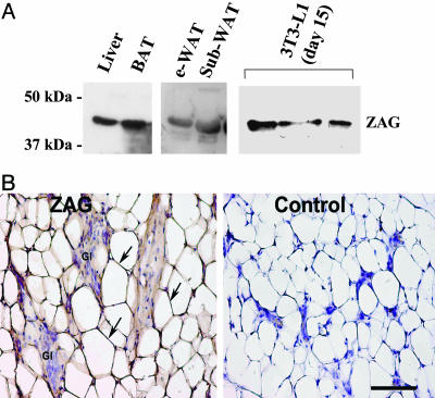
Fig. 3.
ZAG protein in mouse adipose tissue. (A) Western blots of ZAG in liver, brown and white fat, and 3T3-L1 adipocytes (day 15 after differentiation). e-WAT, epididymal WAT; sub, s.c.; BAT, interscapular brown adipose tissue. (B) Immunohistochemical detection of ZAG in mammary adipose tissue. The cytoplasmic rim of white adipocytes shows ZAG immunoreactivity, stained brown (arrows). Gl, glands; internal negative control. (Bar, 75 μmin A and B.)

