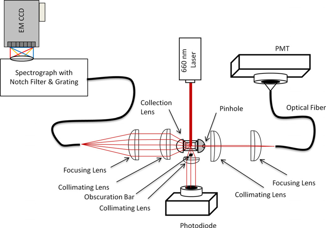
Fig. 2.
Schematic of a Raman flow cytometer. Excitation is provided by a solid state laser (660 nm, 400 mW) and forward angle light scatter is collected on a photodiode. Ninety degree light scatter is collected from one side of the flow cell via an optical fiber and detected with a photomultiplier tube (PMT). SERS signals are collected from the opposite side of the flow cell into and optical fiber and delivered to an imaging spectrograph coupled to a CCD detector. Particles in the probe volume are detected by forward and side scatter, which trigger the acquisition of individual particle spectra by the CCD.

