
FIG 5 .
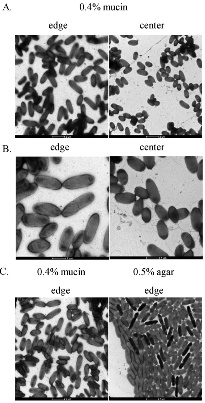
Electron microscopy images of the P. aeruginosa strain PA14 WT from motility colonies on 0.5% agar or 0.4% mucin. P. aeruginosa bacteria were taken directly from the leading edge and center of the mucin-promoted surface motility zone (A and B) or the leading edge of the swarming motility zone (C). The cells were stained with 1% uranyl acetate and observed using a transmission electron microscope.