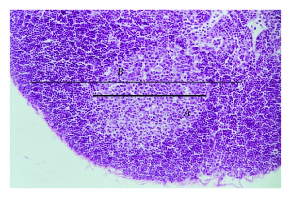
Figure 1.

Photomicrograph of a popliteal lymph node from a thymulin 5CH treated mouse, after 21 days of BCG inoculation. The major axis (B) represents the follicle diameter and the minor axis (A) represents the germinal center. HE staining, magnification 1 : 200.
