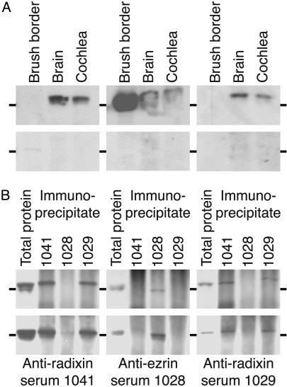
Fig. 1.
Demonstration of the specificity of antisera. Each of the 12 illustrations represents an immunoblot incubated with the antiserum indicated below it. The horizontal marker beside each illustration represents 75 kDa. (A) Peptide competition experiments with anti-radixin and anti-ezrin sera on brush border, brain, and cochlear proteins. (Upper) The anti-radixin sera 1041 and 1029 recognize a protein found in the brain and cochlea, but not in brush borders from intestinal epithelial cells. Anti-ezrin serum 1028 reacts strongly with brush-border protein, moderately with brain protein, and weakly with cochlear protein. (Lower) Probing with antisera in the presence of the corresponding peptides used for immunization eliminates immunoreactivity. (B) Immunoprecipitation and blotting of brain and cochlear proteins to demonstrate the specificity of the anti-radixin and anti-ezrin sera. (Upper) Probing the protein immunoprecipitated from the brain by each of the three antisera demonstrates that the anti-radixin and anti-ezrin sera display negligible cross-reactivity. (Lower) The corresponding experiment for proteins immunopreciptated from the cochlea. In each instance, total brain or cochlear protein is loaded in the first lane as a control.

