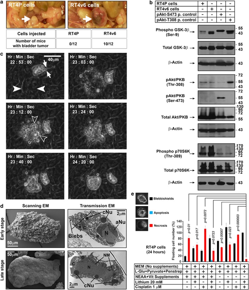
Figure 1.
Inhibition of key signaling pathways that differ in isogenic bladder cancer cell lines RT4P and RT4v6 lead to blebbishield formation. (a) In vivo tumorigenicity of RT4P and RT4v6 cells determined by injecting 200 000 cells orthotopically into the bladder walls of nude mice (n=12 for each cell line). The tumor formation was assessed on day 34 post-injection. Arrow=bladder. (b) Western blotting analysis of regulators of glucose metabolism GSK-3β and Akt, and p70S6K. P. control=positive control; Akt-S473 positive control=UM-UC-3 cells; Akt-T308 positive control=UM-UC-14 cells. (c) RT4P cells at high density (1 × 10⁷ cells/T–75 flask) were exposed to blebbishield ejection (BE) medium and imaged using time-lapse microscopy in DIC bright field settings. Solid arrowheads: the cell(s) starting the blebbishield emergency program. Note a preformed blebbishield in frame 22 : 53 : 00. Arrows in frame 22 : 53 : 00 indicate the thin fragile stalk that anchors the blebbishield with substratum (please also see Supplementary Movie S1). Hollow arrowheads: main body of blebbishield (also see Supplementary Figure S2). Note: The time is counted form the treatment with BE medium. (d) Scanning and transmission electron microscopy of early- and late-stage blebbishields. Note the transmission electron microscopy of early-stage blebbishields with discontinuous cytoplasm. N, nucleus; C, cytoplasm; aNu, active nucleolus; cNu, cytoplasmic nucleolus; NM, nuclear membrane (also see Supplementary Figure S1) (e) Requirement of cisplatin, lithium chloride, and NEAA+Vitamin deprivation to increase the number of blebbishields but to reduce necrosis

