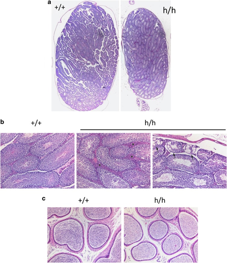
Figure 7.
Impaired spermatogenesis in RNF4-hypomorphic mice. (a) Comparison of testes size in 17-week-old Rnf4 +/+ (left) and Rnf4 h/h (right) male mice. H&E, 12.5x. (b) Comparative microscopic examination of testes in 17-week-old Rnf4 +/+ and Rnf4 h/h (central and right) male mice. Normal testicular histology (left); prominent interstitial compartment with large aggregates of Leydig cells (*) separating seminiferous tubules (central); groups of degenerated seminiferous tubules (arrows) characterized by severe vacuolation of sertoli cells and depletion of germ cells. There was no evidence of tubular degeneration seen in sections from RNF4 +/+ mice. H&E, 100x. (c) Comparative microscopic examination of epididymides in 17-week-old Rnf4 +/+ and Rnf4 h/h males. Normal luminal content with high number of mature spermatozoa was observed in control mice; reduced concentration of spermatozoa associated with degeneration/necrosis of multinucleated spermatids was seen in Rnf4-deficient mice. H&E, 100x

