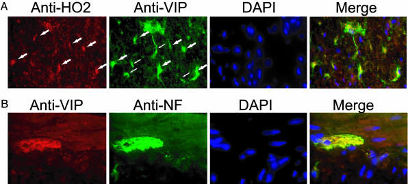
Fig. 1.
Colocalization of HO2 and VIP. (A) Anal sphincter tissue from WT mice was dissected, sectioned, and stained with anti-HO2 (red), anti-VIP (green), and the nuclear stain 4′,6-diamidino-2-phenylindole (DAPI). Neurons of the myenteric plexus in which both HO2 and VIP are present are indicated by large arrows and appear yellow in the merged image. VIP-positive neurons that are not strongly stained with HO2 are indicated by small arrows. (B) VIP (red) is present exclusively in the neuronal innervation of the IAS, as demonstrated by colocalization with the neuronal marker neurofilament (NF; green). The results were obtained in two separate experiments with three mice per group.

