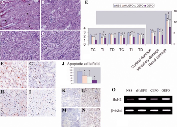
Figure 7.

GEPO ameliorates I/R injuries. Renal histomorphology obtained from NSS (A). rHuEPO (B), CEPO (C) and GEPO (D) groups at 24 h after reperfusion. Severe tubular cell damage was observed in I/R mice receiving NSS treatment (A). Clear necrosis in accompanied by misshapen nuclei in many areas. Improvement of renal histology was remarkably conspicuous after GEPO treatment (D) and also in rHuEPO (B) and CEPO (C) treatment. A semiquantitative scoring system clarified that renal protection resulted from EPO and modified EPO treatment (E) (TC = tubular cast, TI = tubular injuries and TD = tubular dilatation). Notably, GEPO effects were more pronounced for renoprotection, while the histological evidence of CEPO protection was less impressive than that for GEPO and rHuEPO. (*P < 0.05 vs. control), (ŧP = 0.05). A TUNEL assay showed prominent apoptosis of cells in the kidneys of saline-treated animals (F). The numbers of apoptotic cells were significantly decreased in GEPO- (I), rHuEPO- (G) and CEPO-treated (H) mice, as shown by the semiquantitative score in (J). (*P < 0.05 vs. control). Immunohistochemistry analysis demonstrated rHuEPO- (L) and GEPO-treated animals (N) expressed higher Bcl-2 protein in renal tissues as compare with the control (K) and CEPO-treated animal (M). RT-PCR also showed markedly increased intra-renal Bcl-2 transcription in rHuEPO-and GEPO -treated animals as compared with saline-treated animals. Bcl-2 transcription was slightly increased in CEPO-treated mice (O).
