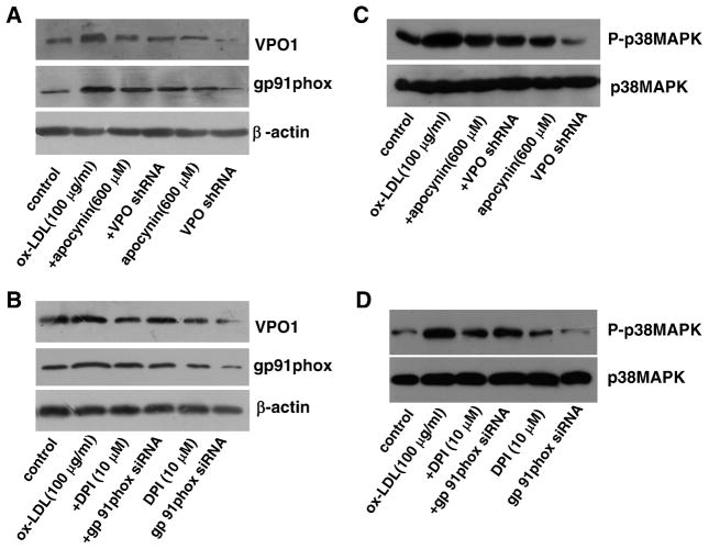
Fig. 5.
Relationship between VPO1 and the NADPH oxidase/p38 MAPK pathway in ox-LDL-induced apoptosis in HUVECs. (A and B) The protein levels of VPO1 and NADPH oxidase subunit gp91phox. (C and D) The protein levels of (phosphorylated) p38 MAPK. Control, endothelial cells were incubated in DMEM containing 1% calf serum for 24 h; ox-LDL (100 μg/ml), endothelial cells were cultured in DMEM containing 100 μg/ml ox-LDL for 24 h; +DPI, cells were pretreated with 10 μM DPI (the specific NADPH oxidase inhibitor) for 1 h before ox-LDL exposure; +apocynin, cells were pretreated with 600 μM apocynin for 1 h before ox-LDL exposure; +gp91phox siRNA, after successful gp91phox siRNA transfection, cells were cultured in DMEM containing 100 μg/ml ox-LDL for 24 h; +VPO shRNA, after successful VPO1 shRNA transfection, cells were cultured in DMEM containing 100 μg/ml ox-LDL for 24 h; gp91phox siRNA, cells with successful gp91phox siRNA transfection were incubated in DMEM containing 1% calf serum for 24 h; VPO shRNA, cells with successful VPO1 shRNA transfection were incubated in DMEM containing 1% calf serum for 24 h; DPI, cells were cultured in DMEM containing 10 μM DPI for 24 h; apocynin, cells were cultured in DMEM containing 600 μM apocynin for 24 h. n=6 each, performed in triplicate.

