
Figure 2. Histological Findings in the Cerebellum.
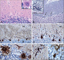
(A) Hematoxylin and eosin: loss of Purkinje cells ; insert, cell with abnormal morphology and unclear outlines of the cytoplasmic membrane. (B) Bielschowski silverstaining: empty baskets (arrows and insert in B). (C–F) Calbindin staining: presence of numerous Purkinje cells with radial sprouting (arrows in C–E and insert in E), reduced and abnormal dendritic arborization of Purkinje cells in the molecular layer (ml; asterisks in D), with swelling of dendritic arborizations (F). (G) Neurofilament staining: swellings of Purkinje cell axons (torpedoes) within the granular layer (gl). Scale bars: A, D: 80 µm; B–C: 160 µm; E–F: 40 µm; G and insert in B: 30 µm; inserts in A and E: 20 µm.