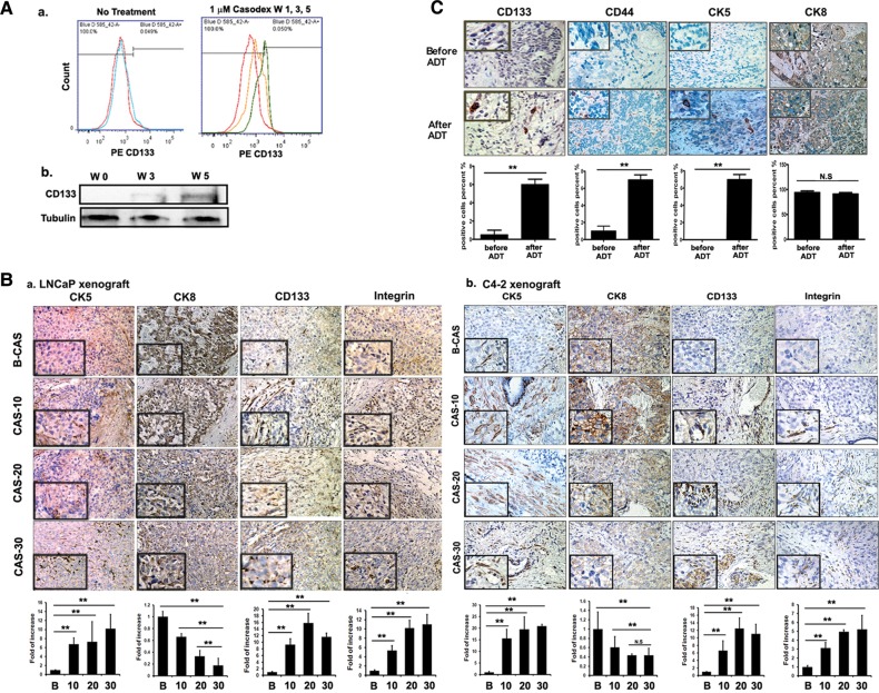
Figure 1.
Stem/progenitor cells increase after castration/ADT. (A) Cell line studies. (a) Flow cytometric analysis of CD133+ cells after 1 (red), 3 (yellow), and 5 (green) weeks of 1 µM Casodex® treatment of LNCaP cells. (b) Western blot analysis of CD133 expression after 5 weeks of Casodex® treatment. (B) Mice tumor tissue studies. Immunohistostaining (IHC) results of tumor tissues. (a) Tumors of LNCaP-xenografted mice and (b) tumor tissues of C4-2-xenografted mice. Tumor tissues were obtained before and 10, 20, and 30 days (indicated as B, 10, 20, and 30) after castration and stained with antibodies of CD133, integrin, CK5, and CK8. Quantitation is shown below the staining data (magnification, ×100; inset, ×400). (C) Human tumor tissues studies. IHC of tumor tissues with antibodies of CD133, CD44, CK5, integrin, and CK8. Human tissues were obtained from Tohoku University Hospital, Sendai, Japan, and Chang Gung Memorial Hospital, Linkou, from the same patients, before and after ADT (magnification, ×100; inset, ×400). **P< 0.01.

