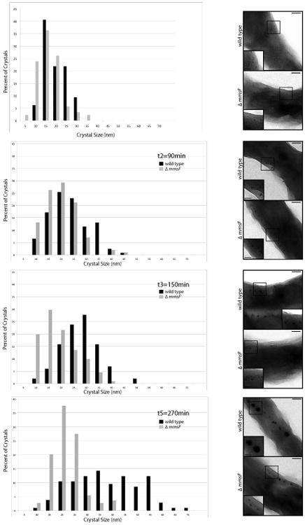
Figure 4. Biomineralization is slowed down and stalls in the ΔmmsF mutant after initial nucleation events.

A. Crystal size distribution in wild type AMB-1 (black bars) and the ΔmmsF (grey bars) strains 30, 90, 150 and 270 minutes post transfer of the cultures in an iron-replete media. The data shown comes from one data set representative of three independent experiments. B. Early stages of magnetite formation in AMB-1 and ΔmmsF. Electron micrographs of glutaraldehyde-fixed cells at the time points indicated in A. Insets are higher magnification of the region boxed in the micrograph. Scale bars: 100 nm, insets 50 nm.
