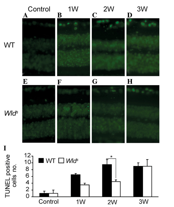
Figure 3.

(A–H) Representative retinal TUNEL staining of the WT and Wlds mice before and 1, 2 and 3 weeks after ON surgery (1:200). (I) Quantitative results of relative number of TUNEL-positive cells in WT and Wlds retina before and 1, 2 and 3 weeks after ON surgery. TUNEL, terminal deoxynucleotidyl transferase (TdT)-mediated dUTP nick end labeling; WT, wild-type; Wlds, Wallerian degeneration slow; ON, optic nerve.
