Figure 5.
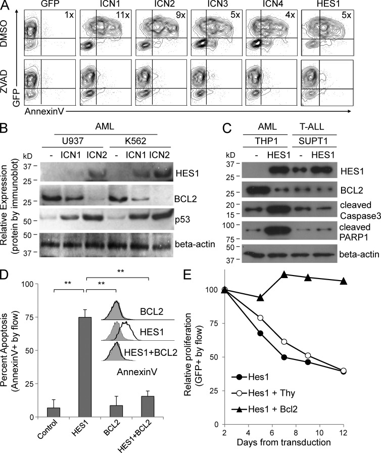
Notch activation induces caspase-mediated apoptosis with BCL2 down-regulation, and HES1-mediated growth inhibition in AML cells is rescued by BCL2 re-expression. (A) Flow cytometry–based contour plots of AML cells transduced with ICN1–4 or HES1 or GFP-only vector and treated with the pan-caspase inhibitor ZVAD, or DMSO vehicle control. Fold increase in Annexin V MFI is noted in each graph (4–11×). Representative of two experiments, and similar results were obtained in the K562 line. (B) Immunoblot of AML cells transduced with GFP vector, ICN1, or ICN2 probed for HES1, BCL2, p53, and β-actin. Representative of two blots. (C) Immunoblot of AML and T-ALL cells transduced with GFP vector or HES1 probed for HES1, BCL2, cleaved caspase 3, cleaved PARP1, and β-actin. Representative of two blots. (D) Flow cytometry–based graph of Annexin V binding percentage in AML cells expressing GFP vector, HES1, BCL2, or HES1 + BCL2 (mean ± SD). Inset shows flow cytometry–based histograms of Annexin V binding from same experiment. Triplicate samples were analyzed and similar results were obtained in an additional cell line. (E) Competitive proliferation assay showing changes in GFP percentage after transduction with HES1, HES1 + Thy vector control, and HES1 + BCL2. Representative of two experiments.

