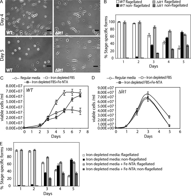
Figure 2.
Iron depletion induces morphological changes and reduced growth rate in WT but not in Δlit1 promastigotes. WT or Δlit1 promastigotes grown to mid-log phase in regular medium were collected and resuspended in iron-depleted medium at 106 cells/ml. Parasites were examined microscopically after staining with the viability dye FDA. (A) Phase-contrast images showing representative WT and Δlit1 parasites on days 4 and 5 during growth in iron-depleted medium. Bars, 12 µm. (B) Quantification of flagellated or nonflagellated WT or Δlit1 parasites at increasing periods after iron deprivation. (C and D) WT (C) or Δlit1 (D) promastigotes grown to mid-log in regular medium were harvested and resuspended in regular or iron-depleted medium or iron-depleted medium supplemented with 8 µM Fe-NTA. Growth was determined by counting viable parasites at the indicated time points. (B–D) The data represent the mean ± SD of triplicate determinations. (E) Quantification of flagellated or nonflagellated WT parasites under the different growth conditions described in C. The data represent the mean ± SD of determinations of three independent experiments.

