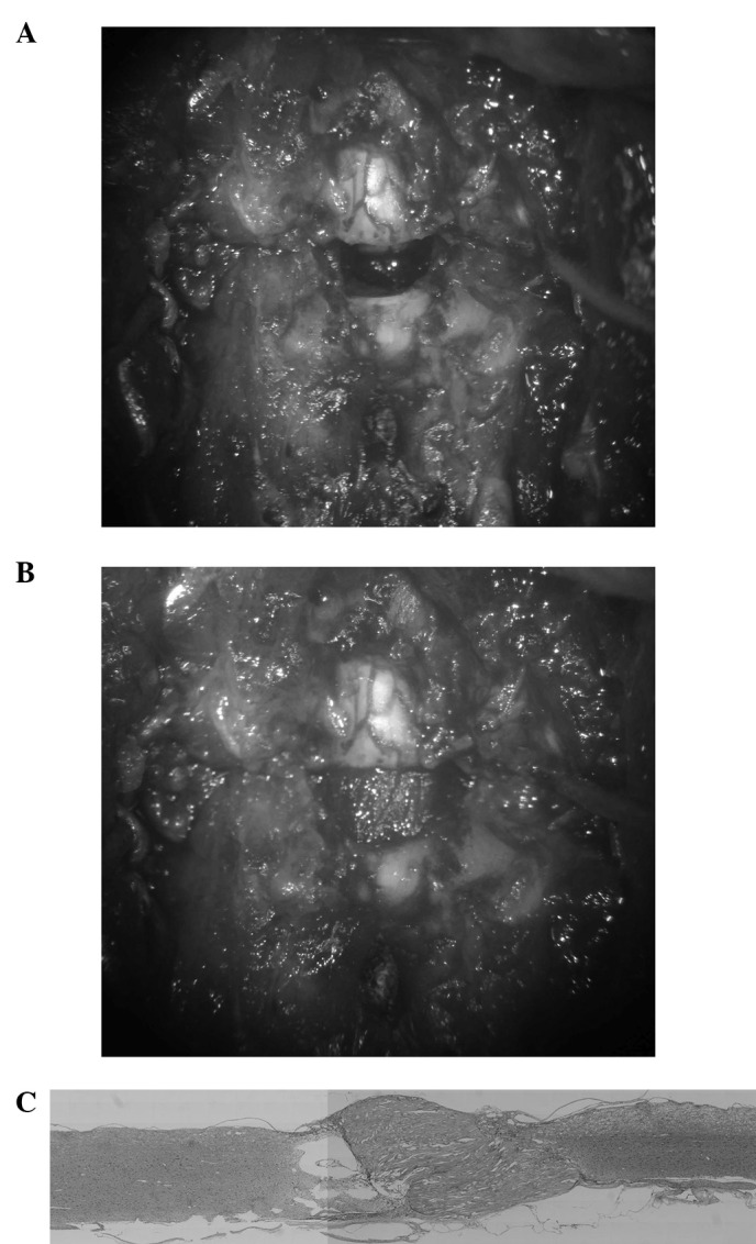
Figure 4.

Scaffold model. (A) Photograph showing the model. A gap of ∼5mm in the spinal cord after shortening of the spine. (B) A scaffold of almost the same size as the resected portion was then implanted in the gap. (C) Light microscopy of the cord showed that the implant firmly connected the stumps of the spinal cord and scar tissue and cavitation were less than in previous models.
