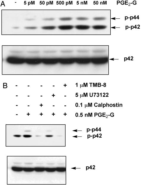
Fig. 6.
PGE2-G induces the activity of ERK in a PKC-dependent manner. RAW264.7 cells were treated for 15 min with vehicle or the indicated concentrations of PGE2-G. Equal amounts (30 μg) of lysate were resolved by SDS/PAGE on 8% gels, and the protein was transferred to poly(vinylidene difluoride) membranes. Membranes were processed for Western blot analysis with an antibody that recognized the phosphorylated forms of p44 (ERK1) and p42 (ERK2) (Upper). Membranes were stripped and reprobed with an antibody that bound modified and unmodified forms of p42. Arrows indicate relative mobility of phospho-p42 and phospho-p44 or p42. (A) Cells received PGE2-G within a concentration range of 5 pM to 50 nM. (B) Cells were pretreated for 15 min with 0.1 μM calphostin, 5 μM U73122, or 1 μM TMB-8 before a 15-min incubation with 0.5 nM PGE2-G. The results shown are from a typical experiment.

