
Figure 2.
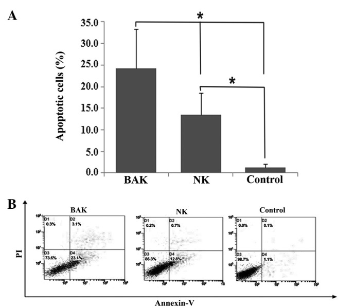
Apoptotic rates of HeLa cells following incubation with BAK and NK cells. HeLa cells without treatment were used as a blank control. Early and late apoptotic cells were combined to calculate the percentage of cell apoptosis. (A) BAK cells had a significant impact on apoptosis in HeLa cells with 24.2±9.2% apoptotic cells compared with the NK control (13.45±5.1%) and the blank control (1.25±0.8%). The NK group also showed significant apoptosis compared with the blank control (*P<0.05). (B) Cell apoptosis was evaluated by using FITC Annexin V/propidium iodide-double staining, and the stained HeLa cells were analyzed by fluorescent-activated cell sorting (FACS). The figure shown is representative of a set of experiments. BAK, BCG-activated killer; NK, natural killer; PI, propidium iodide; BCG, Bacillus Calmette-Guerin.