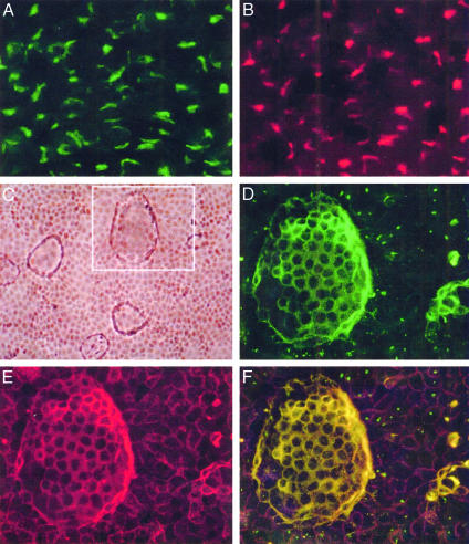
Fig. 1.
rat8 protein localization detection with immunofluorescent microscopy. Undifferentiated and DMSO-differentiated LA7 cells were stained with a polyclonal rabbit anti-rat8 antibody, a monoclonal mouse anti-α6β1-integrin antibody, and a monoclonal mouse antibody against the GM130 protein. (A) Staining for rat8 protein in undifferentiated LA7 cells showing a predominant intracytoplasmic distribution. (B) Staining of the same cells with the GM130 antibody, a Golgi body marker. (C) Phase contrast of DMSO-induced dome-forming LA7 cells. (D) A magnification of Inset in C, which shows a dome-structure stained with anti-rat8 antibody. (E) Staining of the same dome structure with a monoclonal mouse anti-α6β1-integrin antibody. (F) Merged image of D and E. Cells were microscopically photographed with a ×60 objective in A and B, a ×10 objective in C, and a ×40 objective in D–F.

