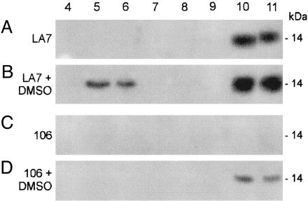
Fig. 2.
Sucrose-gradient fraction analysis for rat8 protein. Western blot analysis of sucrose fractions collected from density gradients of untreated and DMSO-induced LA7 and 106 cells probed with polyclonal anti-rat8 antibody. Equal volumes from each 1-ml fraction, collected as described in Materials and Methods, were separated by SDS/PAGE (12.5% acrylamide) and probed by immunoblotting with polyclonal anti-rat8 antibody. Lane numbers correspond to fractions collected from top (fraction 1) to bottom (fraction 11) of the gradients. Fraction 5 contained a light-scattering band located at the interface between 5% and 30% sucrose. The major part of cells' sphingolipids was associated with this fraction. Fractions 9–11, containing 42.5% sucrose, represent the “loading zone” of these bottom-loaded flotation gradients and contain the bulk of cellular membrane and cytosolic proteins. Fractions 1–3 were omitted because no proteins were normally detected in these fractions of the gradients. Lipid raft-containing fractions correspond to lanes 5 and 6. Lanes 10 and 11 correspond to high-density fractions.

