Fig. 1.
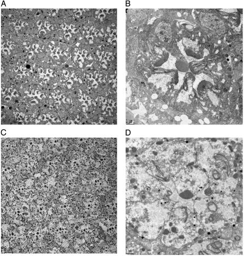
Expression of ceramidase clears rhabdomeric remnants in ninaEI17 mutant photoreceptors. (A) ninaEI17 mutants lacking Rh1 in R1–R6 photoreceptors have defects in rhabdomere biogenesis. A cross section of a compound eye is shown. In transmission electron microscopy sections of 3-day-old fly eyes, all ommatidial units display degenerative rhabdomeres in R1–R6 photoreceptors, and rhabdomeric membranes are visible in the photoreceptor cell body. (Scale bar, 5 μm.) (B) A higher-magnification view across a single ommatidium of 3-day-old ninaEI17 shows that rhabdomeres are incompletely formed, and rhabdomeric elements are seen involuting into the photoreceptors in R1–R6 cells. (Scale bar, 1 μm.) (C) A low-magnification view of 3-day-old ninaEI17 flies expressing ceramidase. In this section, R1–R6 photoreceptors in most ommatidial units have reduced or no rhabdomere, and discrete tubulovesicular structures appear intracellularly. (Scale bar, 5 μm.) (D) A high-magnification view of one of the ommatidial units from C shows that most of the involuting rhabdomeric membranes are missing in R1–R6 cells. Some membranous structures in the cell are seen connected to the apical plasma membrane. Distinct tubulovesicular structures are also seen in the cell body. (Scale bar, 1 μm.)

