Fig. 2.
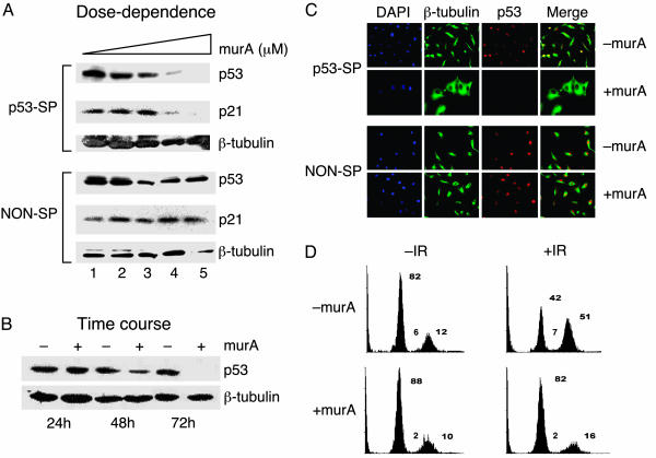
Stable and efficient RNAi-mediated inducible suppression of human p53 gene in U87MG cells. (A) Dose–response of ecdysone-inducible RNAi. Stable cell lines carrying p53-specific shRNA (p53-SP) and nonspecific shRNA (NON-SP) were induced with 0.0 (lane 1), 0.5 (lane 2), 2.0 (lane 3), 3.0 (lane 4), and 5.0 (lane 5) μM murA. Whole-cell extracts were prepared after 72 h and analyzed by Western blotting for p53, p21, and β-tubulin (control). Fold reduction in p53 protein level is 30% (lane 2), 64% (lane 3), 90% (lane 4), and >95% (lane 5) relative to uninduced sample (lane 1). (B) Time course of ecdysone-inducible RNAi. Stable cells carrying shRNA for p53 were induced with 5 μM murA and analyzed for p53 levels at indicated time points; fold reduction of p53 protein level is 60% (+murA at 48 h) and >95% (+murA at 96 h). Fold reduction of protein level was based on densitometric measurement. (C) Inducible suppression of p53 at a single-cell level by immunofluorescence, showing silencing of p53 gene by an inducible p53 shRNA in the presence of 5 μM murA (Upper, bottom row) but not in the absence of murA (Upper, top row), 72 h after induction. Staining with p53-specific antibody (red) and α-tubulin (green). 4′,6-Diamidino-2-phenylindole (DAPI) was used to stain nuclei (blue). (D) Cell cycle analysis of γ-irradiated U87MG cells carrying inducible p53 shRNA by FACS. Stable cell lines carrying p53 shRNA were either uninduced (–murA) or induced (+murA) with 5 μM murA for 72 h and subjected to 20 Gy of γ-irradiation. After 24 h FACS analysis showing distribution of cells in G1, S, and G2/M phases of the cell cycle is shown. IR, irradiation. This experiment is a representation of three independent data sets.

