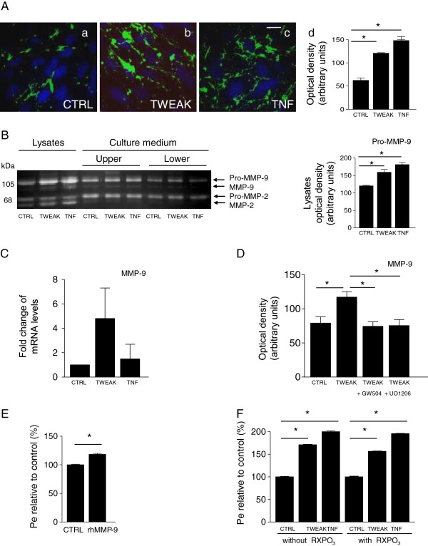
Figure 5.
Gelatinase activity and expression in hCMEC/D3 cells treated with TWEAK and effects of MMP inhibition. (A) Epifluorescence photomicrographs in live hCMEC/D3 cells with gelatin-quenched fluorescent substrate (green), and nuclear intercalant Hoechst (blue). Net gelatinolytic activity (a) increased with TWEAK (b) and TNF (c), shown by densitometric analysis (d). Scale bar, 10 μm. (B) Gel zymography showing expression and secretion of MMP-2 and MMP-9 in cell lysates and culture media after treatment of hCMEC/D3 with TWEAK or TNF. Nonstimulated endothelial cells express pro-MMP-9, pro-MMP-2, and active MMP-2. Active MMP-9 is barely detected. Control cells secrete pro-MMP-9 and pro-MMP-2 in both compartments, and low levels of their active forms. In cell lysates, TWEAK or TNF treatment has no significant effect on the expression of pro- and active MMP-2 but significantly induces pro-MMP-9 and active MMP-9 expression; there is no effect on the secreted forms of MMP-2 and MMP-9. (Densitometric analysis of pro-MMP-9 zymograms of the lysates.) (C) MMP-9 mRNA quantification by qPCR, expressed as fold change ratios of treated versus control samples after normalization with Abelsson mRNA. (D) Inhibition of TWEAK-induced MMP-9 activity by ERK and JUNK inhibitors in hCMEC/D3 cells. After serum-starvation for 24 h, cells were pretreated with ERK and JUNK inhibitors (5 μM GW5074 and 0.5 μM U0126, respectively) for 1 h and then treated with TWEAK. TWEAK readily induces MMP-9 expression. Both inhibitors significantly inhibit TWEAK-induction of MMP-9 in the cell lysate (densitometric analysis of zymograms). (E) Exogenous recombinant human MMP-9 (rhMMP-9, 250 ng/ml for 24 h) enhanced the permeability (Pe) of the endothelial cell monolayer by 20%. (F) Inhibition of MMP activity with the broad-spectrum MMP inhibitor RXPO3 for 24 h has no effect on TWEAK- or TNFα-increased Pe and barrier impairment. In (A,B,D-F) * P < 0.05 according to Student’s t test.

