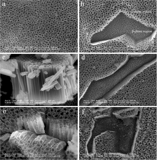
Figure 2.

SEM images of the oxide nanofilms before and after annealing. (a) Top view of as-anodized sample, (b) α- and β-phase regions of as-anodized sample, (c) cross-sectional view of the Ti-A-V-O nanotubes, (d) top view of oxide nanofilms annealed at 450°C, (e) cross-sectional view of oxide nanofilms annealed at 450°C, and (f) top view of oxide nanofilms annealed at 550°C.
