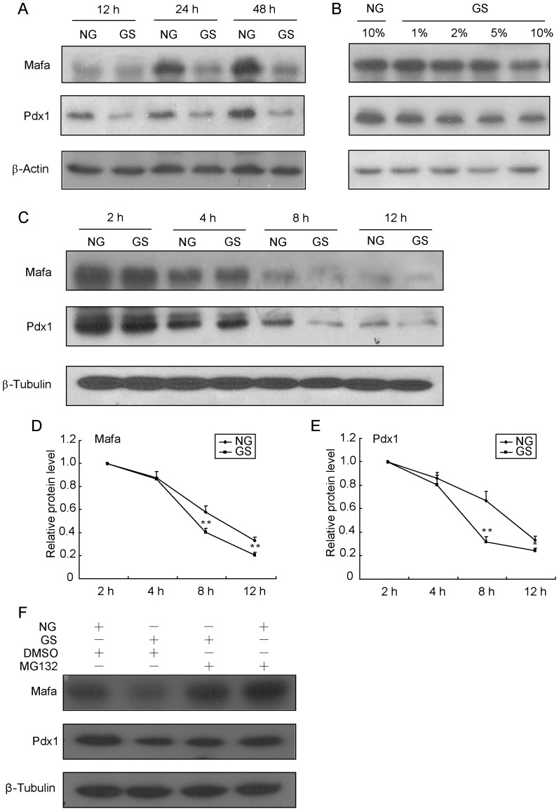
Figure 2. GS induces instability of Pdx1 and Mafa proteins.
INS-1 cells were treated with 10% GS for the indicated times (A) or with different GS concentrations for 16 h (B), and the protein levels of Mafa and Pdx1 were determined by Western blot analysis. (C) INS-1 cells were pre-treated with 50 µmol/l cycloheximide for 2 h and then co-treated with NG or 10% GS for 2, 4, 8 or 12 h. Total proteins were extracted and analyzed by Western blot analysis. (D) Pdx1 and Mafa protein levels normalized to β-Tubulin were compared with the 2 h protein level. (F) INS-1 cells were treated with NG and 10% GS for 12 h, and then MG132 or DMSO was added for an additional 4 h before proteins were harvested to perform Western blot analysis. Values are the mean ± SEM of three individual experiments. *P<0.05 vs. NG; **P<0.01 vs. NG.

