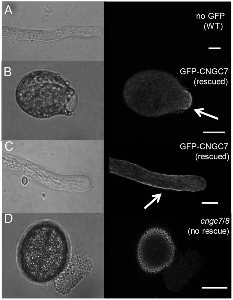
Figure 5. Confocal microscopy showing PM localization for GFP-CNGC7, and the cngc7/8 bursting defect.
Pollen were germinated in vitro and imaged. DIC images are shown to the left, and corresponding confocal fluorescence micrographs to the right. A) A negative control showing a wild type pollen tube without any GFP. B and C), GFP-CNGC7 in cngc7-3−/−, 8-1−/− showing a tip focused PM (plasma membrane) localization at the emerging tube (B) and the tip shank during tube extension (C). D) A non-rescued pollen from cngc7-3−/−, 8-1−/− (segregating a GFP-CNGC7) showing a typical bursting event at germination. Scale bar = 10 µm.

