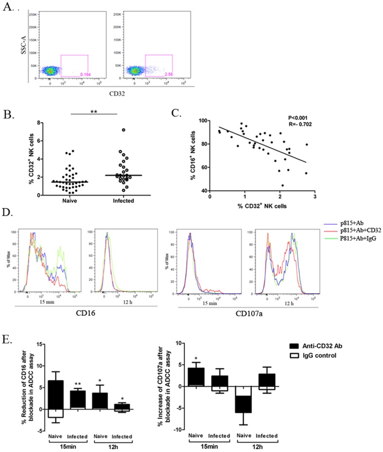
Figure 4. CD32 expression on the NK surface in the naive and SIV/SHIV infected rhesus and its impact on NK cell-mediated ADCC in macaques.
(A) The flow plots represent the CD32 expression on macaque NK cells (right) with the isotype-negative control (left). (B)The comparison of CD32 expression on NK cells from the 40 naive macaques and 23 infected macaques was analyzed. Horizontal bars indicate medians. Mann-Whitney U-test; **P<.01. (C) Correlation between expression of CD16 and CD32 on macaque NK cells is illustrated. Spearman Rank Order Correlation coefficients ‘R’ and corresponding ‘P values’ are indicated. (D) Representative histogram overlays show the expression of CD16 and CD107a on a healthy macaque NK cells activated by Fc targets at 15 min and 12 h in the presence or absence of CD32 blocking antibodies. Irrelevant murine IgG was used as negative control in the blockade assay. (E) The bars represent the reduction of CD16 and increase of CD107a expression on NK cells from 12 healthy and 6 infected macaques in ADCC response after CD32 blockade at 15 min and 12 h. Irrelevant murine IgG was used as negative control. Data represent mean ± SEM. Student paired t test;*P<0.05; **P<.01.

