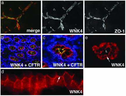
Fig. 3.
Expression of WNK4 in brain, colon, and skin. Frozen sections of mouse brain, human colon, and human skin were stained with anti-WNK4 (red), ZO-1 (green in a), CFTR (green in b and c), or 4′,6-diamidino-2-phenylindole (DAPI) (blue) and analyzed by immunofluorescence light microscopy. (a) Section of brain reveals WNK4 expression is highest in endothelial cells that comprise the blood–brain barrier and overlaps with ZO-1. (b) A low-power view of colonic crypts in cross section, demonstrating WNK4 localizes to the epithelium of crypts. CFTR staining highlights the crypt apical membrane. DAPI staining highlights nuclei. (c) A high-power view of a crypt in cross section showing WNK4, CFTR, and DAPI staining. (d) A longitudinal section of a crypt under high-power demonstrates the localization of WNK4 at intercellular junctions (arrow) in this epithelium. (e) High-power view of a sweat duct in cross section demonstrates that WNK4 is expressed in cuboidal epithelial cells lining sweat duct lumens (L). The arrow demonstrates WNK4's localization along the lateral membrane between adjacent epithelial cells. (Magnifications: ×600, a, c, and e; ×400, b; and ×1,000, d.)

