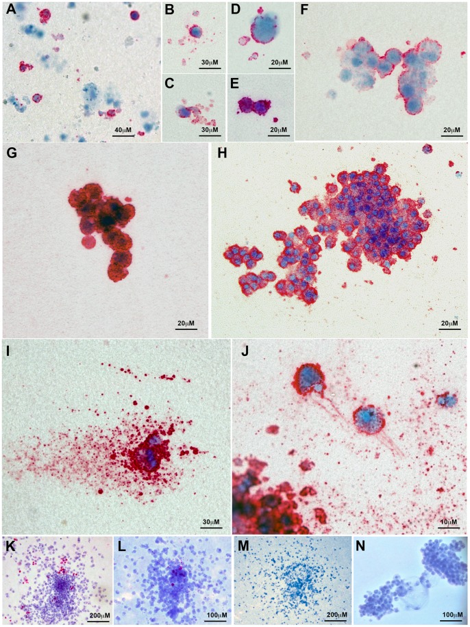
Figure 4. Images of megakaryocytic colonies expressing CD41 cultured from differentiated human ES cells.
Images of colonies were taken from both day 13 and 20 assays. (A) Image showing both CD41-positive and -negative cells within the culture. (B–E) CD41+ cells surrounded by CD41+ cellular fragments, possibly representing platelets. Small (F) weakly and (G) strongly staining CD41+ colonies. (H) Large CD41+ colony (I) Single polyploid CD41+ cell generating many platelet like fragments (J) Section of a Mk colony showing two CD41+ cells and many platelet like fragments, (K–L) Mixed colonies comprising predominantly non megakaryocytic cells (M, N) Two CD41-negative colonies.

