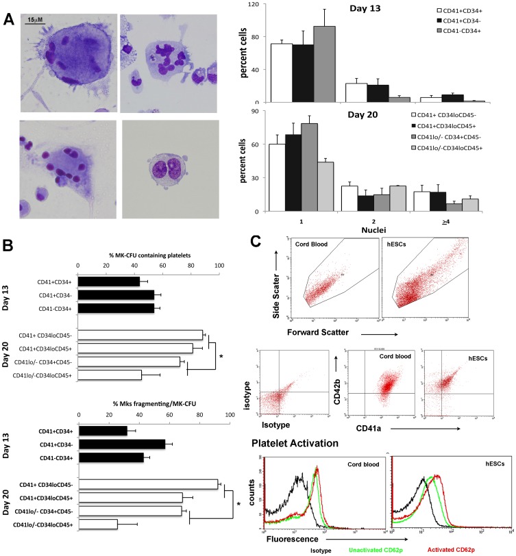
Figure 5. Platelet-like particles generated from human embryonic stem cells (hESCs).
A) May-Grunwald-Giemsa staining of megakaryocytes (Mk) showing endoreplication and membrane blebbing after 13 and 20 days of differentiation of hESCs. The frequency of cells containing 1, 2 or ≥4 nucleii was quantified from stained preparations of single cells at day 13 and 20 of differentiation and results are shown as mean±SEM of n = 4–8 experiments. B) Mk colonies (CFU-Mk) were generated after 13 and 20 days of differentiation by placing differentiated cells in Megacult. The numbers of platelet-like particles were quantitated per CD41+ Mk colony. The upper histogram shows the percentage of CFU-Mks that produced platelet-like particles whilst the lower histogram depicts the percentage of Mks generating platelet-like particles per CD41+ colony. Results shown represent the mean±SEM of n = 4–8 experiments. *, p≤0.05 between the CD41+ sorted fractions and CD41−/lo sorted fractions generated in the day 20 differentiated cells. C) Flow cytometric analysis of platelet-like particles generated from cord blood and hESCs gated on forward scatter and side scatter, showing that the platelet-like particles expressed both CD41a and CD42b, similar to cord blood derived platelets. The lower histogram shows increased expression of the activation marker CD62p in response to incubation with 20 uM of ADP (data from one of two similar experiments is shown).

