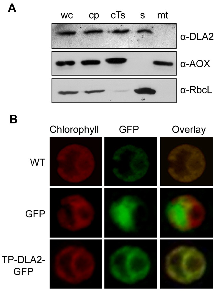
Figure 3. Plastid localization of DLA2.
(A) Cell subfractionation. Cell fractions were prepared as described in Materials and Methods. We separated 30 µg of each protein fraction by SDS-PAGE and subjected them to immunoblot analysis. The blot was probed with antibodies against DLA2 (α-DLA2), the large subunit of Rubisco (α-RbcL), and the mitochondrial AOX (α-AOX, Agrisera). cp, chloroplasts; cTs, crude thylakoids; mt, mitochondria; s, stroma; wc, whole cells. (B) Analysis of the accumulation of DLA2–GFP fusion proteins in the transformed algal UVM4 strain by laser-scanning microscopy. Chlorophyll autofluorescence (Chlorophyll) and expression of GFP without (GFP) or with an N-terminal fusion of the first 114 aa of DLA2 (TP–DLA2–GFP). The untransformed UVM recipient strain served as control (WT). A merged image of the chlorophyll autofluorescence and GFP signals is shown (Overlay).

