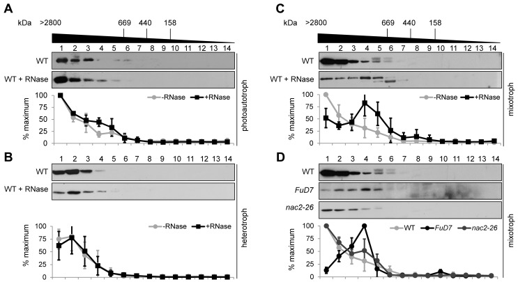
Figure 4. DLA2 forms part of a HMW complex that contains the psbA mRNA.
SEC analyses of the DLA2 complex of wild-type CC-406 cells grown under photoautotrophic (A), heterotrophic (B), or mixotrophic (C) conditions along with PSII mutants FuD7 and nac2–26 grown under mixotrophic conditions. (D) Solubilized crude thylakoid proteins (treated with RNase or not) were separated by SEC. Fractions 1 to 14 were subjected to protein gel blot analyses using the DLA2 antiserum. Molecular masses shown at the top were estimated by parallel analysis of high molecular mass calibration markers. Below each panel, a quantitation of DLA2 signal intensities on Western blots for each condition is presented. The quantitation of signals was performed by using AlphaEaseFC software (Alpha Innotech Corp.). For each experiment, the highest amount of DLA2 in the SEC fractions was set to 100%. Mean values and error bars were calculated from at least three independent experiments.

