Fig. 2.
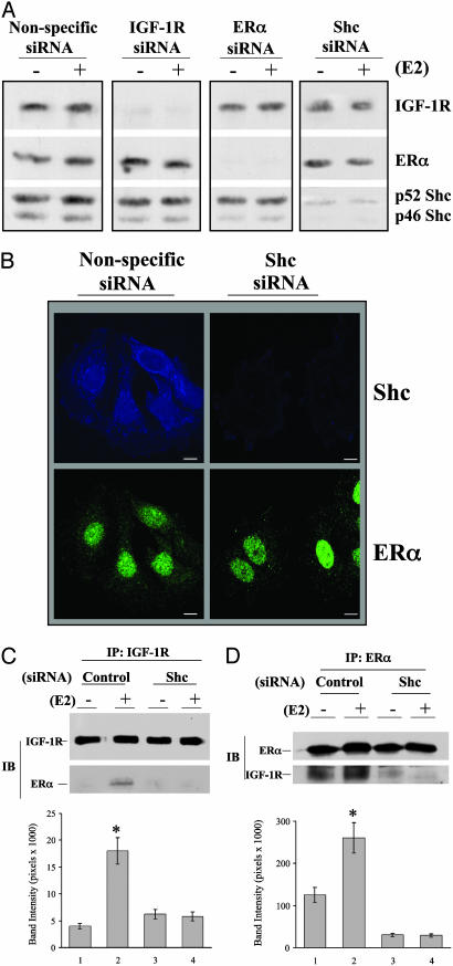
Protein silencing and the role of Shc in protein complex formation in MCF-7 cells. (A) Cells were transfected with nonspecific or specific siRNA for 2 days and then treated with vehicle or 0.1 nM E2 for 15 min for additional MAPK phosphorylation assay. Polyvinylidene difluoride membranes were probed with the specific antibodies to detect the protein expression of IGF-1R, ERα, and Shc. (B) Confocal microscopy study of siRNA expression in MCF-7 cells. Cells, transfected with or without siRNA against Shc, were grown on cover slips and then subjected to immunofluorescence staining for Shc (blue) and ERα (green), as described in Materials and Methods. (C and D) The role of Shc in mediating the interaction of ERα and IGF-1R. Cells transfected with siRNA as indicated were treated with vehicle or E2 at 0.1 nM. The IGF-1R and ERα interaction was assayed by immunoprecipitation of IGF-1R and detection of ERα on Western blot (C) or by immunoprecipitation of ERα and detection of IGF-1R on Western blot (D). The data are from three experiments combined, and only one experiment is shown. *, P < 0.05 comparison of E2-treated cells with vehicle control.

