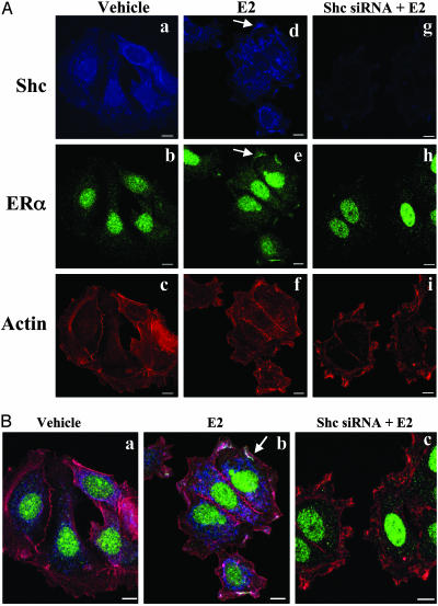
Fig. 3.
Shc involvement in E2-induced ERα membrane association in MCF-7 cells. Cells were transfected with or without siRNA directed against Shc. Two days later, cells were treated with vehicle or E2 (0.1 nM) for 15 min and then subjected to immunofluorescence staining with anti-Shc anti-ERα antibodies. Actin was stained with phalloidin, indicating filamentous cell membrane. (A) The nonmerged images show Shc in blue, ERα in green, and cell membrane in red. (Left) The vehicle-treated cells stained with Shc, ERα, and actin. (Center and Right) Estrogen-treated cells with or without siRNA of Shc expression. (B) Colocalization of ERα, Shc, and actin on cell membrane is shown in three color-merged images. To highlight the specific changes, the arrows illustrate one example of the effects in single cells. Close inspection reveals multiple changes not marked by arrows.

