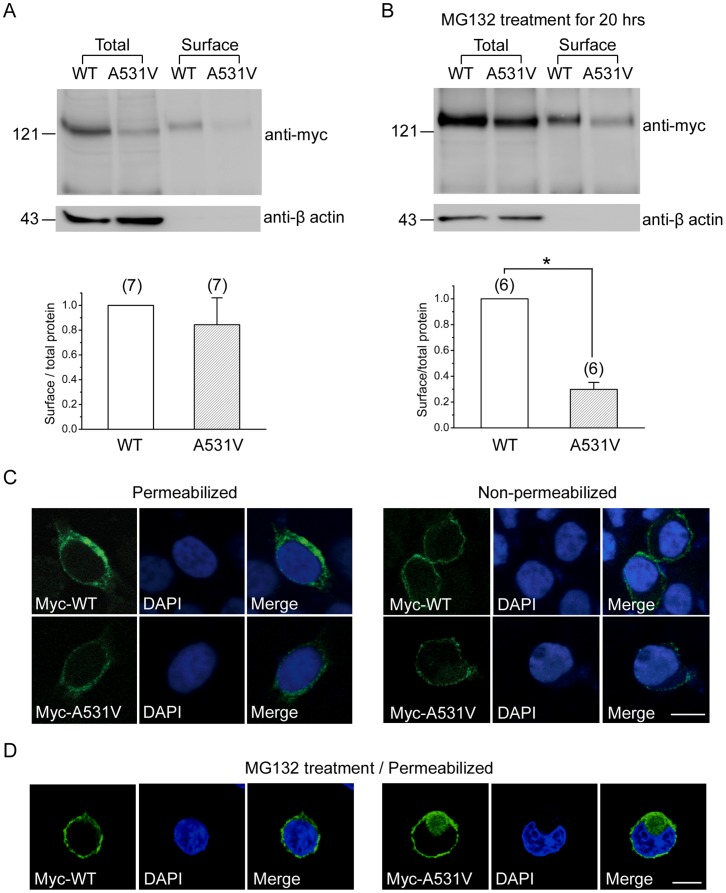
Figure 6. Surface expression efficiency of WT and A531V channels.
Surface biotinylation experiments on HEK293T cells expressing myc-tagged CLC-1 channels in the absence ( A ) or presence ( B ) of 24-hr treatment with 20 µM MG132. (Total) Cell lysates were directly employed for immunoblotting analyses. (Surface) Cell lysates were from biotinylated intact cells, after pulling down with streptavidin beads. To quantify the surface expression efficiency (lower panels), the total protein density was standardized as the ratio of input signal to β-actin signal. The efficiency of surface presentation was expressed as surface protein density divided by the corresponding standardized total protein density. The mean surface expression ratio of the A531V mutant was normalized to that of WT. Densitometric scans of immunoblots were obtained from six to seven independent experiments. ( C,D ) Confocal microscopic images of HEK293T cells expressing myc-tagged CLC-1 channels in the absence ( C ) or presence ( D ) of the MG132 treatment. Fixed cells were stained with the anti-myc antibody (left panels) as well as the nuclear counterstain DAPI (middle panels) under the permeabilized or non-permeabilized configuration. Scale bar = 10 µm.

