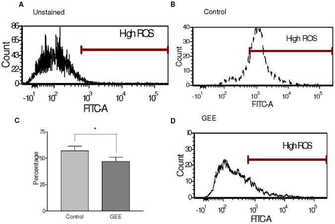
Figure 1. Intracellular reactive oxygen species after islet isolation.
There is a greater percentage of increased fluorescence intensity (High ROS) measured by the excitation and emission of carboxy-h2DCFDA at 495/529 nm (FITC channel), in the control mouse islets (B) when compared to the GEE treated islets (D). ROS content in the control cells (56.9+/−4.33) is significantly higher than in the treated cells (46.98+/−3.94) (p<0.005) (C). Panel A is depicting unstained islets. N = 7 islet isolations.

