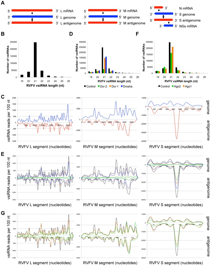
Figure 3. Both the genomic and antigenomic RNA strands of the arbovirus RVFV generate vsiRNAs.
(A) RNA species produced during RVFV infection. (−) strand genomic segments and mRNAs are depicted in blue, (+) strand antigenomes and mRNAs in red. (B) RVFV vsiRNA size distribution (control library). (C) Distribution of 21 nt RVFV vsiRNAs across the three viral genomic segments. vsiRNAs mapping to genomic strand are depicted in blue, antigenomic strand in red. (D) RVFV vsiRNA size distribution between libraries depleted of RNase III enzymes. (E) Effect of RNase III enzyme depletion on 21 nt RVFV vsiRNAs. vsiRNAs from control (black), Dcr-1 (orange), Dcr-2 (green) and Drosha (blue) depleted cells are compared. (F) RVFV vsiRNA size distribution between libraries depleted of Argonaute proteins. (G) Effect of Argonaute depletion on 21 nt RVFV vsiRNAs. vsiRNAs from control (black), Ago1 (orange), and Ago2 (green) depleted cells are compared. See also Figures S1, S2.

