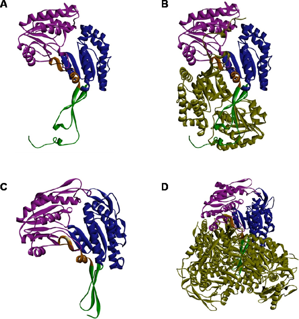
Fig. 2. Protein structure of ALDH3A1 and ALDH1A1.
X-ray crystal structures of human ALDH3A1 monomer (A) and sheep liver ALDH1A1 monomer (C) showing the catalytic domain (purple), NAD(P)+ binding domain (blue), and oligomerization domain (green). Crystal structures were obtained from RCSB database; PDBIDs: 3SZA (ALDH3A1) and 1BXS (ALDH1A1). Computational modeling of dimeric human ALDH3A1 holoenzyme (B) and tetrameric sheep ALDH1A1 holoenzyme (D). The 2nd ALDH3A1 subunit and three other ALDH1A1 subunits are shown in forest green.

