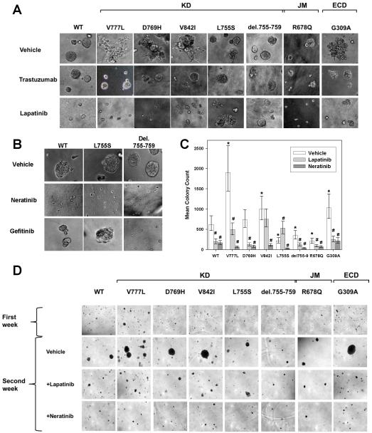
Figure 4.
HER2 mutations V777L, D769H, V842I, G309A induce gain of function over HER2 WT in MCF10A mammary epithelial cells. A, HER2 WT or mutants were seeded on 3D Matrigel culture in the presence or absence of DMSO vehicle (0.5%), trastuzumab (100 μg/ml) or lapatinib (0.5 μM). Phase contrast images of acini or invasive structures were obtained at 200x magnification on day 8. KD = kinase domain, JM = juxtamembrane region, and ECD = extracellular domain. B, HER2 WT, L755S, and del.755-759 cells were grown in Matrigel in the presence of DMSO vehicle (0.5%), neratinib (0.5 μM) or gefitinib (0.5 μM). Phase contrast images were obtained as in A. C, MCF10A-HER2 WT or mutants were seeded in soft agar. After 7 days of growth, they were treated with DMSO vehicle (0.5%), lapatinib (0.5μM) or neratinib (0.5μM) for an additional week. Error bars represent 95% highest posterior density intervals. * indicates a significant difference between the HER2 mutant and HER2 WT and # indicates the effect of inhibitor treatment was significant (95% highest posterior density interval did not contain 0 for both). D, Photomicrographs of the colonies in soft agar on day 12, magnification 40x.

